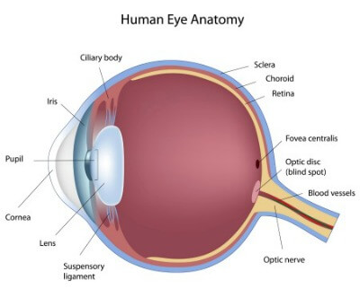
 Our Austin retina specialist deals primarily with conditions that affect the posterior aspects of the eye. Most frequently this involves the vitreous gel, retina and the choroid.
Vitreous humor:
This structure is a gel filling the cavity of the eye between the lens and the retina at the back of the eye. This gel can cause visual problems in its interface with the retina. It can cause pulling on the retina leading to tears. In addition the cavity it occupies can fill with blood in certain conditions.
Retina:
This is a thin structure that lines the posterior wall of the eye. This “wallpaper” of the posterior eye is nerve tissue that collects light rays and images and passes them to the brain through the optic nerve enabling vision. Conditions disrupting the retina can cause visual difficulty.
Choroid:
This is a vascular structure behind the retina supplying oxygen to the posterior section of the eye. This structure can be involved in conditions causing bleeding or leaking fluid beneath the retina.
Age related macular degeneration:
Age-Related Macular Degeneration (ARMD) is the leading cause of vision loss in older American adults. You may hear ARMD referred to as being either dry or wet. Typically, dry ARMD is the initial type of change that occurs in susceptible eyes. Dry changes can be detected on a dilated retinal exam. These changes can cause mild to significant visual difficulty. In general dry ARMD progresses slowly. However, the changes in dry ARMD may allow defects in the choroid that allows new and abnormal blood vessels to grow under the retina. These blood vessels may leak or bleed. When this occurs the condition becomes wet ARMD.
Currently there are no treatments for dry ARMD. However, you may be placed on vitamins to help slow progression. You will likely be asked to monitor your vision with an Amsler grid, which you may find on this web-site. There are currently several medications used to treat wet ARMD and new medications are being researched. You treatment options will be discussed during your exam.
Diabetic retinopathy:
Among the many adverse health effects of diabetes is it happens to be one of the leading causes of blindness in the United States. Diabetes has many varied effects on the eye. Visual loss can occur from swelling in the retina from leaking blood vessels. Abnormal blood vessels may grow in response to poor oxygenation in the eye. These vessels may break and cause bleeding in the eye that fills the vitreous space and decreases vision. These vessels can also contract and lead to retinal detachment. Treatment may involve surgery, laser or injection of medication.
Retinal vein and artery occlusion:
Occlusions of blood vessels in the eye cause loss of vision by loss of blood flow to retina tissue or by swelling caused by leakage from damaged vessels. People with high blood pressure and diabetes are at increased risk of experiencing this condition. Loss of vision due to inhibited blood flow is not treatable. However the visual loss from swelling is treatable with intraocular injection of medication or laser. Exam and eye imaging techniques help to determine if treatment will be beneficial.
Flashes and floaters:
These are common symptoms and will frequently pass over a period of time. The issue is that the symptoms may indicate a retina tear or retina detachment. The only way to assure that these serious conditions are not present is to perform a dilated eye exam.
Retinal tears and detachment:
Retina tears occur when there is a separation of the vitreous gel from the retina at the back of the eye. This separation is a normal process in aging. It usually occurs without causing a tear or detachment. However, the symptoms of flashes or floaters commonly occur. If a tear is found during an exam it may be treated with laser. If it has progressed to a retina detachment a surgical procedure will likely be necessary.
Macular hole:
The macula is the name given to the central part of the retina. A macular hole occurs here and effects central vision and reading. This condition is treated with a surgical procedure that involves removing traction from the retina. A gas bubble is left in the eye and recovery requires a period of keeping your head in a face down position.
Epiretinal membrane:
A membrane is a type of scar tissue that grows over the central retina or macula. This tissue contracts and distorts the retina decreasing the quality of central vision. It is removed with a surgical procedure called a vitrecotmy.
Schedule an appointment with our Austin Retina Specialist today!
